Preclinical Imaging

One Instrument, Many Applications

Our line of compact MRI systems is designed to facilitate a wide variety of in vivo applications and research models, empowering scientists, pharmaceutical and research professionals to explore and address a myriad complex biological challenges.

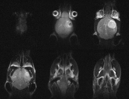
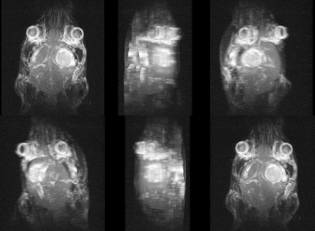
Braintumor-in-vivo-1
Brain-tumor-in-vivo2-1

Our systems are designed to be used to routinely and repeatedly quantify phenotypes in models of disease, including:

  • Cancer
  • Cardiovascular
  • Contrast-based Molecular Imaging
  • Diabetes and Obesity
  • Embryology/Developmental Biology
  • Nephrology
  • Neurobiology
  • Stem Cell/Cell Tracking

Anatomy and Morphology

In vivo soft tissue imaging for morphological characterization. 2D and 3D imaging can be performed quickly and easily for preclinical model assessment.

C_Anatomy-and-Morphology

Cancer Research

Detection, follow-up, and quantification of tumor development and progression.

D_Cancer-Reasearch

Neurobiology

In vivo anatomical imaging of the brain, spine and spinal cord for assessment and follow-up of neurologically-based diseases.

E_Neurobiology

Multi-modality Imaging

Easy registration with other modalities such as Optical, PET, SPECT and CT to enable powerful multi-modality phenotyping.

F_Multi-modality-imaging

Anatomy and Morphology

In vivo soft tissue imaging for morphological characterization. 2D and 3D imaging can be performed quickly and easily for preclinical model assessment.

G_Molecular-Agent

Ex Vivo Imaging

High-resolution, high throughput, 3D MR-based histology imaging of fixed samples and embryos for toxicological and developmental studies.

Ex-Vivo-Imaging

Anatomy and Morphology

  • Respiration/cardiac triggering
  • Preceding inversion recording pulse
  • Diffusion weighted imaging

Anatomy and Morphology

  • 2D and 3D
  • Respiration/cardiac triggering
  • Dynamic acquisition – i.e. Dynamic Contrast Enhanced (DCE)
  • IR Snap for T1 map generation

Anatomy and Morphology

  • 2D and 3D Respiration/cardiac triggering variable echo train length
  • Multi-point fat/water separation

Anatomy and Morphology

  • Respiration, ECG and temperature monitoring
  • Respiration and ECG output triggering to MRI spectrometer

Anatomy and Morphology

  • Vaporizer with temperature and flowrate compensation
  • Scavenging cube for waste gas
  • Breathing circuit with 3 access points
Need for Information?

Speak with a specialist today to learn more about Aspect Imaging’s full suite of compact, high-performance MRI systems and preclinical imaging applications.

Aspect Imaging
Privacy Overview

This website uses cookies so that we can provide you with the best user experience possible. Cookie information is stored in your browser and performs functions such as recognizing you when you return to our website and helping our team to understand which sections of the website you find most interesting and useful.

To view our full privacy policy, please visit this link: https://aspectimaging.guybenami.com/privacy-policy/